Active Nano Mapping Facility


PI: Dr Neil Fox
Funded through the NNUF2 round, the Active Nano Mapping (ANM) Facility based at the University of Bristol, hosts the world’s fastest atomic force microscope (AFM), a high-speed AFM (HS-AFM). The microscope is housed in a dedicated active lab capable of handling samples up to a contact dose of 50 µSv/hr (or higher if the safety case can be made). Full glovebox facility enables the processing and imaging of samples without exposure to ambient conditions.
About the microscope
HS-AFM is a Bristol Nano Dynamics Ltd. contact mode AFM that can collect video rate topography maps of surfaces with sub-atomic height and nanometre lateral resolution in ambient, controlled gas, and controlled liquid environments. The system has significantly higher throughput when compared to a conventional AFM, being able to collect and process a year’s worth of AFM data in a matter of hours. This can be used in three main ways:
https://www.youtube.com/embed/yQivN39XdVI
Video shows real-time data collection, left shows a conventional AFM, right shows HS-AFM. Each frame is 2 microns wide.
Copyright University of Bristol, 2019-2020 all rights reserved
1. To observe dynamic nano and micro scale processes in controlled liquid, gaseous, inert or ambient environments.
Easy-to-use navigation software and the ability to image under in-vivo conditions allows the ANM Facility to observe processes such as corrosion, oxidation, or cracking with sub nanometre resolution and frame rates up to 100 frames per second. The imaging window can be easily moved around the sample to find structures of interest. It is often the case that the nanoscale structures of interest are not homogeneously distributed on a surface and that most of the challenge in their measurement is in locating them in the first place. HS-AFM has proven itself to be the perfect tool for this task. To aid in this process optical images are collected simultaneously with topography and stored with the metadata.
Video shows the dissolution of zeolite in de-ionised water.
Copyright University of Bristol, 2019-2020 all rights reserved
2. To map areas millimetres in size with nanometre resolution.

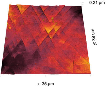
Dislocations on a steel surface
Reprinted from International Journal of Fatigue, 127, Payam, A. F., Payton, O., Picco, L., Moore, S., Martin, T., Warren, A. D., Mahmoud M., and Knowles, D., 'Development of fatigue testing system for in-situ observation of stainless steel 316 by HS-AFM & SEM', 5, Copyright (2019), with permission from Elsevier.
The HS-AFM can make use of the high frame rate by moving between frames to build up a composite image. This allows areas up to millimetres in size to be imaged without a reduction in resolution.
3. To collect spatial maps of the distribution of the physical dimensions of nanostructures with world-leading statistical validity over millimetre sized areas.

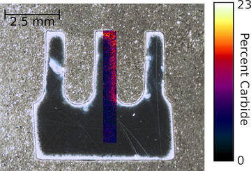
The distribution of carbides in a cooling fin as measured by HS-AFM and processed on the NanoMapper software package.
Copyright University of Bristol, 2019-2020 all rights reserved. Data from Liu et al.
The imaging window can be moved across the sample surface in an automatic fashion in order to collect a sparse data set of thousands of frames in a matter of minutes. Custom software then characterises over 80 physical properties of every identified nanostructure on the surface. The processing can be filtered to only measure the nanostructures of interest. The spatial distribution of the nanostructure of interest can then be mapped on top of a calibrated macro image to enable a complete understanding of the macro scale distribution of nano and micro scale structures. The example shown here maps the carbide content in a 9 chrome steel boiler component. This process could also, for example, give you the distribution of mean diameter for all nanostructures orientated at between 30 and 50 degrees and below 2 microns in size.
Imaging modes
The system has the following capabilities:
- Dynamic strain up to 2kN
- Heated sample stage up to 200oC
- Image in ambient, liquid, gas environments with controllable humidity
- Ability to exchange liquid and gas environments while imaging
- Ability to load samples within an inert glovebox environment
- Sub-atomic height resolution, ~1 nm lateral resolution
- Topography, electrical, thermal and stiffness imaging modes
- Macro and micro optical images.
Please contact us for more information so that we might help you plan your experiments using the active HS-AFM in the ANM NNUF at the University of Bristol.
Please email stacy.moore@bristol.ac.uk or loren.picco@bristol.ac.uk for more information.

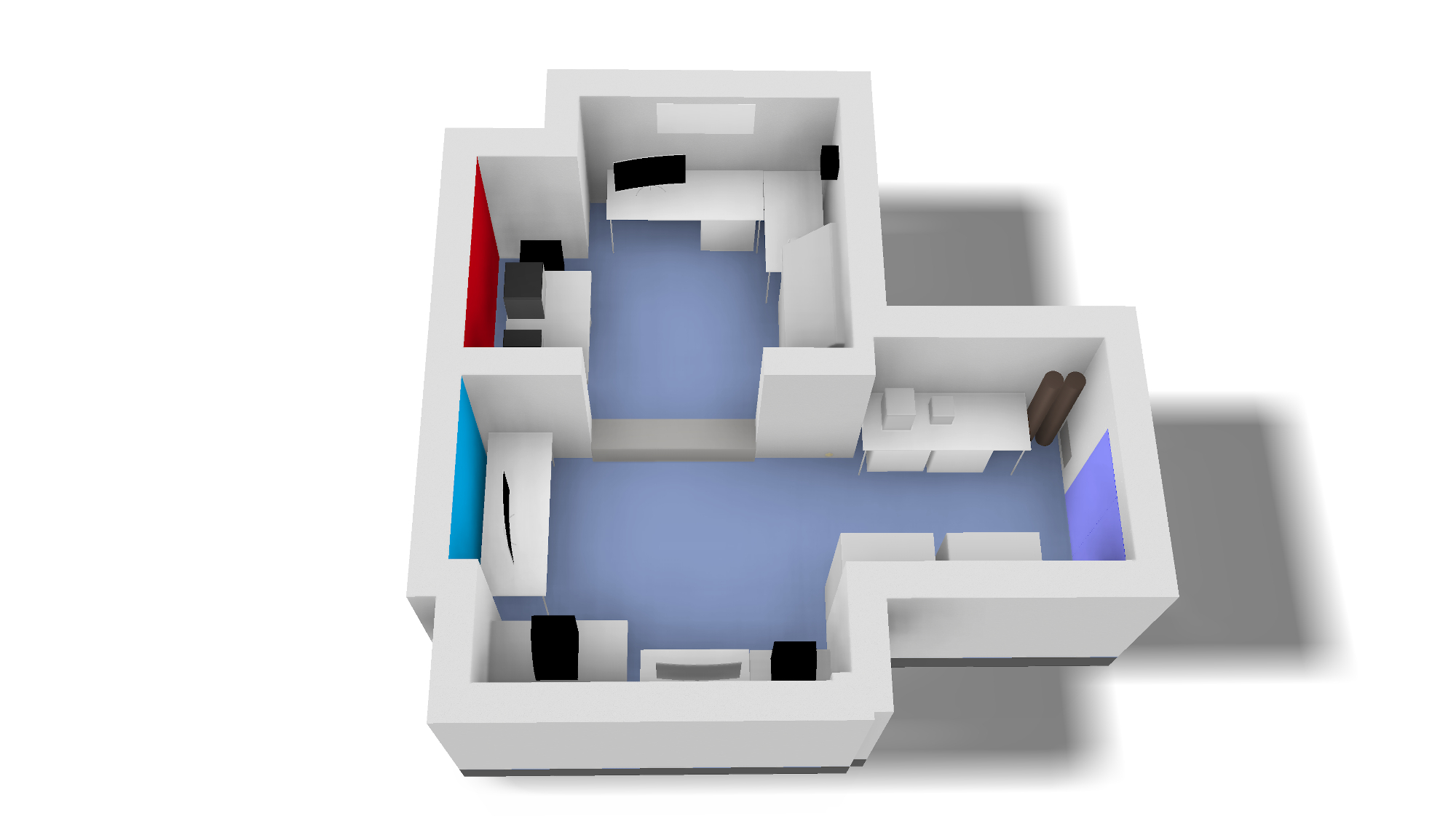
A render of the ANM NNUF Laboratory at the University of Bristol showing the active (red) and non-active (blue) sections.
Copyright University of Bristol, 2019-2020 all rights reserved
Availability
The ANM Facility is open for research.
Copyright University of Bristol, 2019-2020 all rights reserved.


